New Technology Makes Waves in Iowa State Animal Science Lab
Iowa State University students are using cutting-edge 3D anatomy visualization to see livestock in a new light.
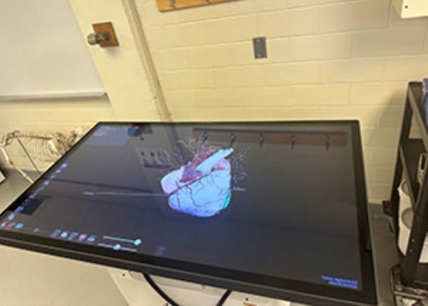
Karl Kerns, an assistant professor of animal science at Iowa State University, is opening students' eyes in a unique way in a lab that accompanies the animal science 214 lecture class. The lab provides students with detailed examination of organs and biological systems of domestic animals.
“The learning tool is called the Anatomage Virtual Dissection Table,” Kerns said in an Iowa Pork Industry Center release. “It is preloaded with hundreds of labeled anatomical system images, as well as the ability to read native MRI or CT Scan files which makes it invaluable for topics like the circulatory system and the skeletal system.”
The technology allows students to see images that replicate what it looks like in a living species. There's no question that seeing blood move throughout the heart and body in real time, with the valves opening and closing, gives students a different perspective from a traditional specimen.
The table can lie flat like an actual table and it can also stand like a monitor to fit different learning circumstances, the release said. It has a touch screen surface for interactive learning and the entire assembly is on wheels, allowing it to move from classroom to classroom.
Kerns said the device has proved to be an invaluable learning mechanism with just a few months into its use. It offers an alternative tool for students unable to participate in traditional methods, such as those with allergies, he noted.
Read More:
9 Questions Pork Industry Leaders Challenge You to Think About
Scott Dee Announces Retirement; Reflects on Top 10 Lessons Learned
Legacy Means Different Things To Different People: Pig Farmers Respond