The Bull Lameness Exam
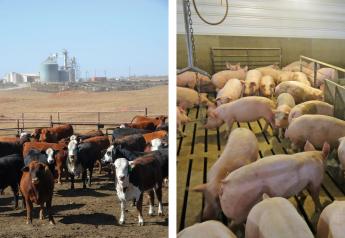
The bull lameness exam Lameness can mean the difference between a valuable bull and hamburger. Identifying lameness and correctly diagnosing the cause of bull lameness can result in treatment success and potentially the retention of a valuable animal.
Meredyth Jones, DVM, MS, Dipl. ACVIM, Kansas State University, says 90% of lameness cases can be isolated to the foot, with 90% of claw lesions in the hind feet, mostly in the lateral claw. The causes of lameness in bulls are many (see sidebar), so it’s important to take a targeted approach to diagnosis.
Footrot is the most common cause of lameness in cattle, but most of those cases are managed on-farm. “The most common things I see on farms as primary care cases are toe abscesses and subsolar abscesses,” Jones says. “I also see a fair number of subcutaneous abscesses that occur over the shoulder or thigh, usually as the result of fighting. Beyond those, spinal trauma, developmental orthopedic diseases such as osteochondritis dissecans and spastic paresis occur with enough regularity that I keep an eye out for them.”
Spastic paresis, spastic syndrome, and corkscrew claw all likely have a heritable component. “We certainly see spastic paresis associated in cattle with overly straight hocks,” she says. “The developmental orthopedic diseases, like osteochondritis dissecans, are multifactorial with genetics, nutrition and environment likely all contributing. The real take home message is that, when making breeding and culling decisions, musculoskeletal and hoof quality should be important considerations.”
Jones offers these recommendations for conducting a lameness exam on a bull.
1. START WITH HISTORY
Jones starts every lameness exam with as complete of a history on the bull as she can get. “Duration of the lameness and any previous history of lameness or systemic illness prior to this episode are useful to know when evaluating the animal,” Jones says. “Of particular interest to me when I’m evaluating a case is the progression of this episode of lameness, treatments that have been done on the farm and any response to those treatments.”
Jones says one of the most common things she sees is that the bull becomes lame, he gets treated with antimicrobials, usually oxytetracycline because it’s presumed to be footrot, and then he doesn’t respond. “At this point, the bull needs to be examined because a simple case of footrot will almost always respond to a single dose of oxytetracycline,” she explains. “When that is the history, the veterinarian knows to go on a more aggressive hunt for the cause of the bull’s lameness.”
2. WHAT’S HIS JOB?
Depending on how the bull is used can influence treatment choice or disposition. The primary career choices for bulls are to be range or pasture bulls, AI sires or a combination. “We generally don’t see bulls in the feedlot, or prefer not to, but this may be a career option for young bulls that perhaps were intended to be sires, but were channeled into the feeder route because of another reason,” Jones says. “Certainly, producers are generally more willing to go for more aggressive therapy with higher economic value bulls. I believe and teach my students that every case you see has three options for management: low-cost, middle-of-the-road and the Cadillac. Each of those has a cost estimate, aftercare and prognosis associated with it and veterinarians are there to educate their clients so they can make informed decisions.”
From another perspective, what the bull is expected to do for a living plays heavily in his prognosis for certain diagnoses. “For example, if the bull has distal interphalangeal septic arthritis in the lateral claw of the hind limb, we may be able to surgically manage him to be an AI sire or for small pasture breeding, but he may have reduced longevity in a large rangeland situation,” Jones says. “We want to be able to be very up front with producers about the prognosis for return to soundness for each type of breeding situation so that they can decide in their own mind if that is acceptable to them.”
3. LOOK AT THE WHOLE PACKAGE
Jones says when she looks at a bull for any reason, his body condition is always something on her mind. “If a young bull presents for lameness and he is very obese, it may be a clue as to his nutritional management (very high energy feeds and overfeeding) and would move developmental orthopedic disease up on my list,” she says.
Jones adds how he got to the body condition he’s in may be a bigger factor in his disease and recovery than his actual condition at the time of evaluation. “Has he been out covering a lot of cows over a lot of ground? Is he still a young, growing bull that is breeding cows and is not being fed to meet both of those demands and he’s just getting run down in general? If he’s overfat, is that extra weight contributing to the strain on his joints or will it make his recovery more difficult? In many cases, body condition may not have a direct impact on lameness, but it certainly adds to the bull’s entire story which must be considered when managing him.”
4. THE “DISTANCE EXAM”
Certainly, the mantra of most veterinarians is that they want the animal caught up when they arrive on the farm, and Jones holds to that as well, but with a slight difference. “For lameness cases, my definition of ‘caught up’ is in a small trap or pen where there is room to walk him around. I don’t need 400 acres to see him walk, but likewise, I cannot evaluate his gait or see his whole body in the alleyway or chute either.”
Jones explains that when she can see the whole bull and walk him, she can look for subtle swellings, figure out exactly which leg is the problem or see if it is her impression that the problem might be up in his spine. “His degree of lameness also gives me a lot of information about the cause of his lameness. If I see a swelling in the area of a joint and the bull won’t put the leg down, then I have a pretty good feeling that the joint is septic, rather than just having a sprain or effusion.”
Jones says while she don’t see them with great regularity, diseases like spastic paresis and spastic syndrome are actually best diagnosed while observing the gait and once they are in the chute, there’s not really much to do with them except rule out other disease processes.
Jones adds that she can also evaluate his symmetry and detect any muscle atrophy by watching him walk. Disuse atrophy generally becomes noticeable after about two weeks of disuse, which helps the veterinarian get a sense of the duration of the lameness.
In cases where animals that are severely lame or have evidence of neurologic disease, Jones has to really consider the wisdom of even putting the animal in the chute and risking not being able to get them out. “In those cases, I’m going to consider casting restraint or sedation in a pen if further evaluation is necessary.”
5. RESTRAINT AND PALPATION
After Jones has seen the bull walk and feels comfortable sending him to the chute, she prefers to palpate the entire limb, particularly when it is a hind limb, prior to using a rope to pick up the foot. “We know that greater than 90% of lameness lesions are in the foot, so it’s logical to go there first, but if this guy has a partially ruptured cruciate ligament and I put a rope on his foot and pick it up and he struggles for a minute, which they always do, then I may have just finished off his cruciate.” The same could be true for fractures or other unstable upper limb lesions.
Many cases have obvious swelling and when that’s the case, Jones palpates for any heat or coolness of the area and determining the character of that swelling: is it soft and fluctuant, firm or hard like bony proliferation? As the swelling increases in firmness, the chronicity of the process is usually increased and will likely result in the need for more aggressive therapy.
From the elbow and stifle up, palpation is more challenging. “I will often go back and forth from one elbow or shoulder to the other comparing the size and character of the joint to convince myself that the joint on the lame leg is normal or abnormal,” Jones suggests. “In the case of the stifles, I am generally able, even on large bulls, to place my hands on both stifles from behind the bull at the same time, making the comparison of size more direct.”
Evaluation of the hip requires palpation of the position of the greater trochanter of the femur relative to the ileal wing and ischiatic tuberosity. The three should form a triangle with the greater trochanter at the low point and if the greater trochanter is displaced , this indicates a hip dislocation. Further evaluation of the hip and pelvis really requires rectal palpation and casting restraint or sedation to put the limb through range of motion to palpate for crepitus.
6. HAVE THE RIGHT TOOLS
Jones carries in the truck a foot box that contains a hoof tester, hoof nippers, half-round hoof nippers, Swiss knife, regular hoof knife, stainless steel teat cannulas, lidocaine, innertube tourniquet and butterfly catheters for foot anesthesia, and supplies to place a wooden block. “The most common hoof issues that I see are toe abscesses and subsolar abscesses. They can be discovered by trimming with foot trimmers or paring away the sole with hoof knives. Once discovered, these need to be opened completely, wide enough to provide good drainage. This is not for the faint at heart and, if I’m going to get down to sensitive tissue, I will place a tourniquet at the mid-cannon bone and perform a Bier block (regional intravenous perfusion of lidocaine) to anesthetize the foot to allow me to be aggressive. As I’m working, I use a teat cannula to probe to find every bit of undermined tissue and get it removed.”
Jones advises to not quit too soon. “Sometimes, I’ve had cases where I removed the entire sole. If it is undermined, it has to go. I have found halfround hoof nippers to be the best tool for peeling off the dead, infected sole.”
Jones finds that ultrasound is useful in evaluating the fluid character of effusive or enlarged joints, and for diagnosing septic tenosynovitis, as a backup to her clinical impression. “People who are really experienced with ultrasound can find cartilage flaps, bursitis and even fractures with ultrasound,” she says. If arthrocentesis is to be performed for cytology, protein concentration and culture of the joint fluid, ultrasound guidance is useful.
Radiographs are also useful in some lameness cases and portable units can provide some good images of lower limb conditions. “All that said, I really believe that the vast majority of cases can be accurately diagnosed, prognosed and treated after a thorough clinical exam.”
For more on bull health and bull examinations, read these articles on BovinVetOnline:
BSEs for Young and Mature Bulls
Sidebar
How bull lameness happens
Bulls are aggressive and frequently groups of bulls are put together, often ranging in age. Young bulls, when placed with an established group of bulls, should be monitored closely to ensure that they don’t become the whipping boys which usually manifest as shoulder injuries, explains Meredyth Jones, DVM, MS, Dipl. ACVIM. “Whenever possible, bulls should be allowed to spread out. Confinement, even for a short time, often results in a fight.”
A bull’s hind legs and back are under a tremendous amount of strain during breeding. “Acute injuries, from mild sprains to ligament tears can occur, but the chronic wear-and-tear is what prematurely ends the careers of most bulls,” Jones says. “Spinal and limb pain may be severe enough to reduce semen quality to an unacceptable level or can result in a bull limiting or even totally ceasing to mount cows.”
Bulls often have to travel long distances such as in range pastures, and footing is a contributor to lameness. “Two environmental conditions that most significantly affect a bull’s feet are very dry and very wet,” Jones says. “Very dry ground predisposes hooves to cracking vertically, which can get deep enough to pinch the sensitive laminae with every step. Very wet ground is a big risk factor for footrot, but can also result in softening of the soles, making them more vulnerable to bruising.”
Causes of lameness
Jones offers this list of common causes of lameness, categorized by which part of the foot or leg they are usually found. “My goal with this is to give veterinarians a starting point of ideas to entertain when they localize a lameness to a particular anatomic location,” Jones explains.
“In general, this can be done fairly easily during the clinical evaluation, but every once in a while, they can be tricky. In lameness evaluation of horses, local anesthesia as a means of lesion localization has become mainstay. We do it some in cattle, but it certainly isn’t common. The disease processes which cause lameness in cattle tend to be infectious or inflammatory, making them fairly easy to spot in most cases.”
Foot: Mechanical causes
• Sole ulcer/sole abscess/toe abscess
• Corkscrew claw
• White line disease/laminitis
• P3 fracture>P2>P1
• Interdigital fibroma (corn)
• Vertical wall crack
• Horizontal crack
Foot: Infectious/septic causes
• Septic arthritis of DIP joint>MTP>PIP
• Septic tenosynovitis
• Pedal osteitis
• Papillomatous digital dermatitits
Metatarsus/Metacarpus
• Physitis
• Fracture
• Cellulitis
Carpus
• Hygroma
• Septic arthritis
Elbow
• Septic arthritis
Humerus
• Hematoma
• Fracture
Shoulder
• Hematoma
• Septic arthritis
• OCD
Hock
• Conformation issues
• Septic arthritis
• OCD
Tibia
• Fracture
• Cellulitis
Stifle
• Septic arthritis
• Cranial cruciate rupture
• OCD
Femur
• Diaphyseal fracture
• Distalphyseal fracture
Hip
• Slipped capital physis
• Coxofemoral luxation
Spine
• Trauma
• Fracture/luxation
• Spastic syndrome
• Ankylosing spondylitis
• Spinal lymphoma
• Epidural abscess
• Vertebral body abscess